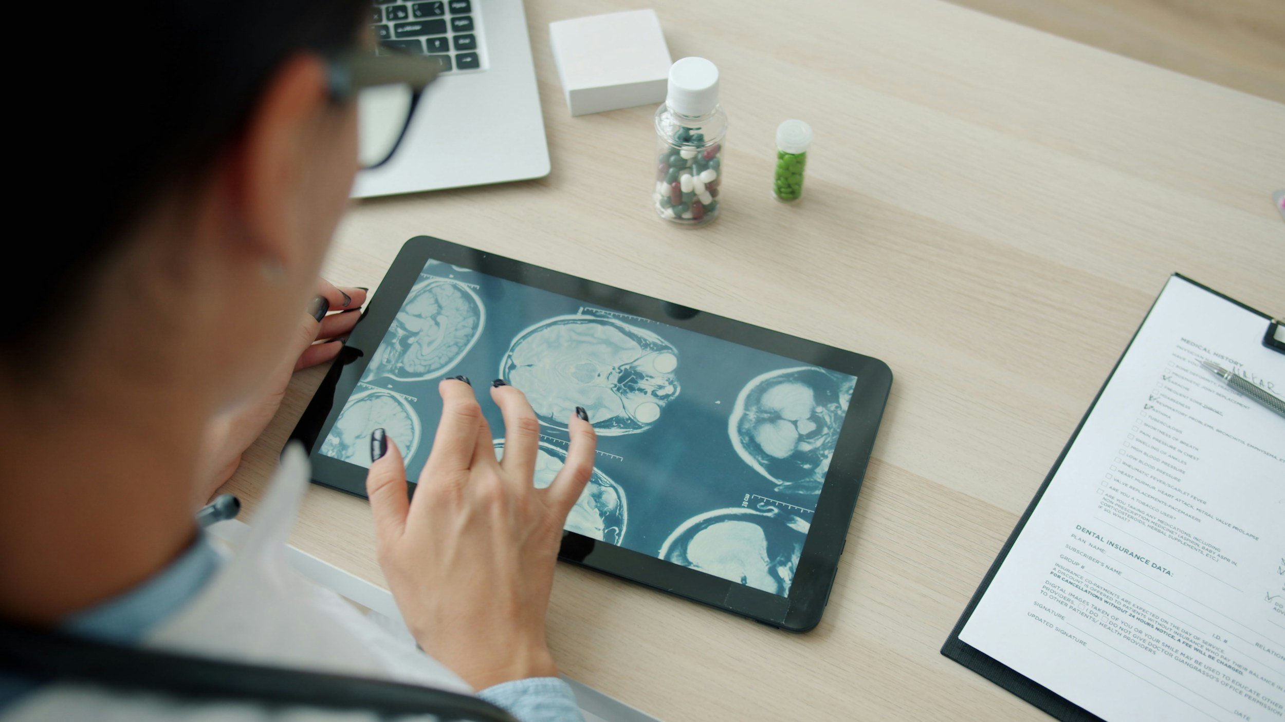
How to Read a Back MRI Report
If you have recently had an MRI scan for back or neck pain, you may be wondering what to expect when you get your results. As with many medical imaging reports, the terminology used to describe the spine can be confusing. This article covers the specific language you might encounter in your results for a lumbar spine MRI or cervical spine MRI and other back MRIs.
Your doctor will provide the medical interpretation of your scan, but understanding terms like MRI views, signal intensity, and common spinal findings can help you have a more enlightened follow-up appointment.
What Does a Spine MRI Show?
Magnetic resonance imaging (MRI) is a non-invasive technique that utilizes radio waves and powerful magnets to create comprehensive images of your internal structures without the use of radiation. A spine MRI provides detailed visualization of the soft tissues, including the spinal cord, nerves, and intervertebral discs. It is a primary tool for diagnosing the root cause of back pain and sciatica.
Common conditions a spine MRI might help diagnose include:
Herniated or bulging discs pressing on nerves.
Spinal stenosis (narrowing of the spinal canal).
Nerve root compression (radiculopathy).
Trauma or injury, such as vertebral fractures.
Infections (e.g., discitis) or tumors.

MRI showing disc herniation
Possible Findings in Spine MRIs
Cyst: A fluid-filled sac, such as a "discal cyst," which is rare but can cause symptoms similar to a herniated disc.
Edema: Inflammation or swelling caused by fluid build-up, often seen in the bone marrow (Modic changes) following injury or severe degeneration.
Lesion: A general term for any tissue abnormality, which could be due to injury, illness, or aging.
Mass or Tumor: Unexpected tissue growths that may be benign (non-cancerous) or malignant.
MRI Views: Axial, Coronal, and Sagittal
To ensure the most accurate diagnosis, MRI images are taken from three different angles, providing a 360° overview of your spine:
Sagittal View: A side view of your spine. This is often the best view to see disc height and how the vertebrae align.
Axial View: A horizontal cross-section, like looking down through a vertebra. This view is crucial for seeing if a disc is pinching a nerve root.
Coronal View: A frontal view, presenting a mirror image of your spine from front to back.
T1 vs. T2 on MRI: What is the Difference?
A radiologist uses different "weightings" to highlight different tissues.
T1-weighted MRI: Best for showing the anatomy of the spine. Fat appears bright (high signal), while water and cerebrospinal fluid (CSF) appear dark.
T2-weighted MRI: Highly sensitive to pathology because it highlights water and fluid. On a T2 scan, healthy, hydrated discs and CSF appear bright.
Understanding Signal Intensity
Signal intensity refers to the level of brightness in an image.
Low T1 Signal: If bony areas appear darker than normal, it may suggest a decrease in healthy fatty marrow, potentially indicating infection, trauma, or lesions.
High T2 Signal: Areas that appear "whiter" than surrounding tissue often indicate increased water content, such as swelling (edema), cysts, or inflammation.
Fluid Signal: These look bright on T2 and dark on T1, helping doctors identify fluid-filled structures like an abscess or a syrinx (a fluid cavity in the spinal cord).
Understanding the Sections of Your MRI Report
Indication: A brief summary of the symptoms or medical reasons (e.g., "lower back pain") that led to the MRI.
Findings: The descriptive section where the radiologist records every observation, noting what is normal, abnormal, or age-related.
Unremarkable: This is good news! It means the area being examined appears normal with no signs of disease or noteworthy abnormalities.
Impression: The most important section. The radiologist summarizes the key findings and provides a potential diagnosis for your doctor to review.
Next Steps: Clinical Correlation
It is common to feel nervous about "abnormal" findings, but findings like "disc desiccation" or "mild bulging" are often normal signs of aging and may not be the cause of your pain. Your specialist will perform a "clinical correlation," matching the images to your physical symptoms to determine the best treatment plan.
By the Brain and Spine Neurosurgical Institute of Rhode Island
Like this content?
We have an educational newsletter allowing you to stay informed on your spine health.